[コンプリート!] venous sinuses of brain mri 196331-Venous sinus thrombosis brain mri
Brain herniations into dural venous sinuses (DVS) are rare findings recently described and their etiology and clinical significance are controversial We describe five patients with brain herniations into the DVS or calvarium identified on MRI, and discuss their imaging findings, possible causes, and relationship to the patient's symptomsCerebral Venous Thrombosis Radiology department of the Medical Centre Haaglanden in the Hague and the Rijnland hospital in Leiderdorp, the Netherlands Cerebral venous thrombosis is an important cause of stroke especially in children and young adults It is more common than previously thought and frequently missed on initialMar 01, 16 · Dural venous sinuses 3 Characteristic feature of dural venous sinuses • Lined by endothelium, no muscular coat & valveless • Collect blood from brain,meninges, orbit,internal ear & diploe • Connected to valveless emissary veins to maintain the internal & external venous pressure • Projection of arachnoid granulation into it for CSF

Focal Brain Herniation Into A Dural Venous Sinus An Incidental Rare Entity
Venous sinus thrombosis brain mri
Venous sinus thrombosis brain mri-Dural venous sinus thrombosis is diagnosed on CT venography and MRV but CT plain brain is the initial radiological investigation Most of the time CT shows signs such as hyper density, hyperdense delta sign On this scan a subtle linear hyperdensity was seen in the region of right transverse sinus and confluence of sinuses6 rows · Dec 11, 16 · The most prevalent type of CVST is dural sinus thrombosis (or sinus thrombosis, SVT), which
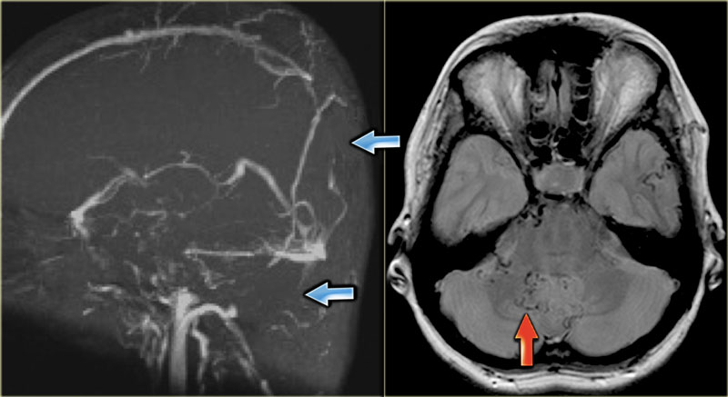


The Radiology Assistant Cerebral Venous Thrombosis
Background and Purpose—In cerebral venous thrombosis (CVT), the sensitivity of conventional MRI sequences to detect clot in the sinuses or veins is incomplete and largely depends on the time elapsed since thrombus formation Little is known concerning the corresponding diagnostic value of fluidattenuated inversion recovery (FLAIR), echoplanar T2*Knowledge of variations in the cerebral venous anatomy and apparent signal abnormalities seen on Magnetic resonance (MR) angiography are essential to avoid overdiagnosis of cerebral venous sinusSep 01, 07 · Venous engorgement with enlargement of the dural venous sinuses, enlargement of the pituitary gland, thickening and subsequent enhancement of the loose connective tissue of the subdural space, and transudative fluid collections within this space, as well as frank subdural hematomas are all phenomena that "compensate" for the loss of intracranial volume
Apr 01, 21 · There were no evidence of cerebral infarction or demyelinating lesions from MRI of the brain, but MR venography showed thrombosis of the terminal part of the superior sagittal sinus, straight sinus, and rightsided transverse and sigmoid sinus Occurrence of cerebral venous sinus thrombosis is extremely rare in scrub typhus, and only two casesTABLE 3 Diagnostic Performance of Brain MRI Sequences for Characterization of Acute Dural Venous Sinus T hrombosis (DVST), According to PerSegment Analysis of 624 Segments For characterization of acute DVST, the 3D T1weighted GRE CE sequence had the highest sensitivity for DVST ( p < 005), compared with all other sequences, although allAbstract Objectives To determine frequency, imaging features and clinical significance of herniations of brain parenchyma into dural venous sinuses (DVS) and/or calvarium found on MRI Methods A total of 6160 brain MRI examinations containing at least one highresolution T1 or T2weighted sequence were retrospectively evaluated to determine the presence of incidental brain
Embryologically, a sinus by that name does exist, collecting the venous drainage of the very thin cortical mantle (future superficial sylvian veins), and directing it from the sphenoid area towards the sigmoid sinus, underneath the temporal lobe This sinus usually disappears in the adult, except for some very rare instancesThe intracranial or cerebral venous system is a network of nerves made up of two systems working together the superficial system and the deep system(1) The superficial cerebral system has sagittal sinuses and cortical veins The sinuses and the veins both drain deoxygenated blood from the surfaces of the brain's hemispheres(2)When MRI of the brain is the initial exam, a T1 hyperintense thrombus or loss of T2 flow void effect may be found within the dural sinus (Figure 2) The gradient echo sequence may demonstrate susceptibility hypointensity within the thrombosed venous sinus (Figure 2C)


Radiological Imaging Of Cerebral Venous Thrombosis Ppt Video Online Download


Cerebral Venous Sinus Thrombosis Secondary To Otomastoiditis Postgraduate Medical Journal
About Press Copyright Contact us Creators Advertise Developers Terms Privacy Policy & Safety How works Test new features Press Copyright Contact us CreatorsApr 13, 21 · Cerebral Venous Sinus Thrombosis (CVST) is a type of rare blood clot that forms in the venous sinuses in your brain This is a system of veins found between the layers ofMRV is used to assess abnormalities in venous drainage of the brain 2dimensional (2D) timeofflight (TOF) MR venography (MRV) and 3dimensional (3D) phasecontrast (PC) are the technique commonly used to assess the cerebral venous sinuses because they are easy to perform and do not require contrast administration
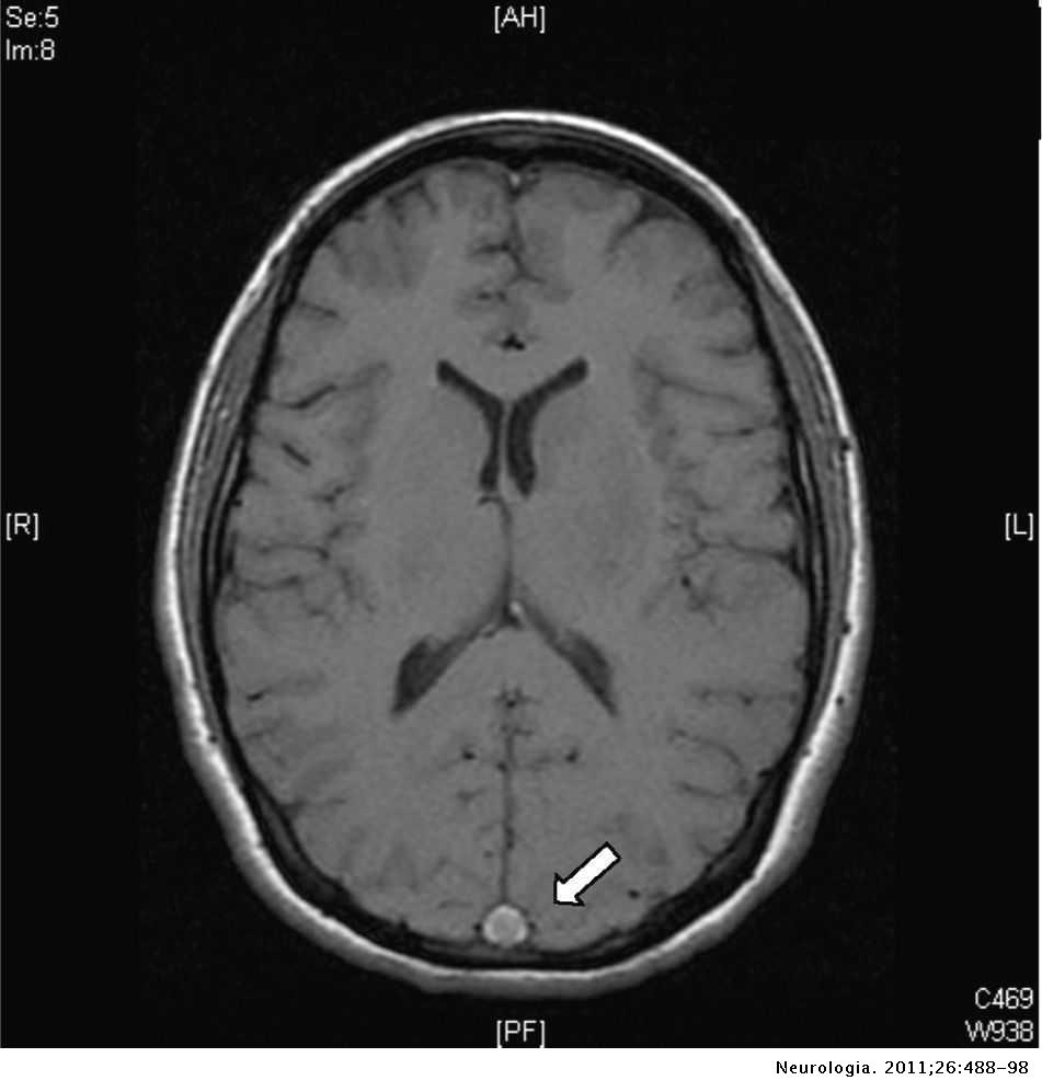


Cerebral Venous Thrombosis A Diagnostic And Treatment Update Neurologia English Edition


Current Endovascular Strategies For Cerebral Venous Thrombosis Report Of The Snis Standards And Guidelines Committee Journal Of Neurointerventional Surgery
Further evaluation with diagnostic techniques like magnetic resonance imaging (MRI) and magnetic resonance (MR) venography is preferred when cerebral venous sinus thrombosis is suspected CT or catheter venography can substitute for MR venography, but MRI is more sensitive than CT in detecting early parenchymal infarctionThrombosis of the sinuses impedes venous drainage from the brain and may result in venous infarction within the region of brain that drains into the sinus This involves the thalami in thrombosis of the straight sinus and vein of Galen ( Fig 914 ) or the cortex and subcortical white matter with thrombosis of the sagittal sinus ( Fig 915 )Cerebral venous sinus thrombosis (CVST) occurs when a blood clot forms in the brain's venous sinuses This prevents blood from draining out of the brain As a result, blood cells may break and leak blood into the brain tissues, forming a hemorrhage This chain of events is part of a stroke that can occur in adults and children


Cerebral Vein And Dural Sinus Thrombosis Intechopen


Cerebral Venous Sinus Thrombosis On Mri A Case Series Analysis
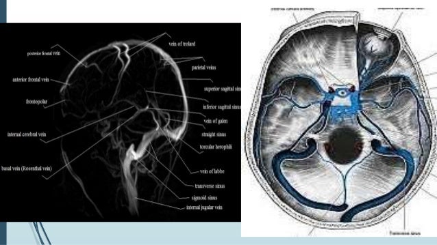
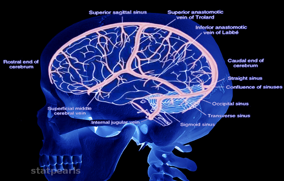
Cerebral venous sinus thrombosis (CVST), cerebral venous and sinus thrombosis or cerebral venous thrombosis (CVT), is the presence of a blood clot in the dural venous sinuses (which drain blood from the brain), the cerebral veins, or bothOne of the causes of acute neurological deterioration is cerebral venous sinus thrombosis (CVST) It is a rare form of stroke and results from thrombosis of dural venous sinus (which drain blood from brain) Intracranial dural sinus thrombosis is potentially fatal and relatively common conditionOct 21, 10 · Cerebral venous thrombosis is located in descending order in the following venous structures Major dural sinuses Superior sagittal sinus, transverse, straight and sigmoid sinuses Cortical veins Vein of Labbe, which drains the temporal lobe Vein of Trolard, which is the largest cortical vein that drains into the superior sagittal sinus


1


Venous Sinuses Neuroangio Org
May 06, 21 · MRI Atlas of the Brain This page presents a comprehensive series of labeled axial, sagittal and coronal images from a normal human brain magnetic resonance imaging exam This MRI brain crosssectional anatomy tool serves as a reference atlas to guide radiologists and researchers in the accurate identification of the brain structuresJul 07, 19 · MRI of brain vessels without contrast Magnetic resonance imaging of cerebral vessels without the use of a contrast agent is carried out to assess the condition of veins and arteries The main indications for diagnosis Stroke (hemorrhagic, ischemic) Aneurysms Thrombosis Vascular pathologyMar 09, 16 · The thrombus occludes venous return through the sinuses, and causes an accumulation of deoxygenated blood within the brain parenchyma This in turn can lead to venous infarction The situation is complicated by an accumulation of cerebrospinal fluid, which can no longer drain through the thrombosed venous sinus
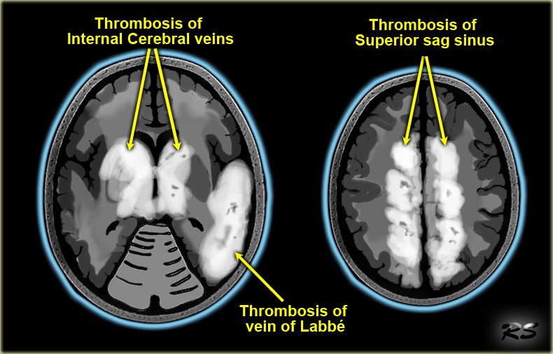


The Radiology Assistant Cerebral Venous Thrombosis


The Radiology Assistant Cerebral Venous Thrombosis
Feb 01, 14 · Brain herniations into dural venous sinuses (DVS) are rare findings recently described and their etiology and clinical significance are controversial We describe five patients with brain herniations into the DVS or calvarium identified on MRI, and discuss their imaging findings, possible causes, and relationship to the patient's symptomsSuspected venous sinus thrombosis Undetermined vascular malformation Venous varyx, Galen vein anomalies MRA HEAD W/O CONTRAST SPECIFY MRV ONLY Please note, these are possible indications Please specify MRV study HEAD AND NECK Skull ENT or Neurosurgical stereotactic approach MRI HEAD W CONTRAST (UMC order appear s as MRI BRAIN WCerebral vein and cerebral venous sinus thrombosis Cerebral vein and cerebral venous sinus thromboses are blood clots that form in the veins that drain the blood from the brain called the sinuses and cerebral veins They can lead to severe headaches, confusion, and strokelike symptoms They may lead to bleeding into the surrounding brain tissues


Brain Magnetic Resonance Imaging Study Showing Cerebral Venous Sinus Download Scientific Diagram
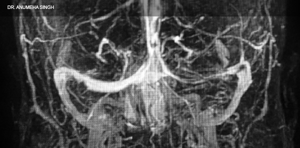


How To Spot And Treat Cerebral Venous Sinus Thrombosis Acep Now
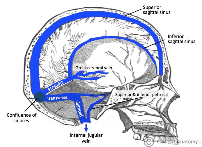
The dural venous sinuses (also called dural sinuses, cerebral sinuses, or cranial sinuses) are venous channels found between the endosteal and meningeal layers of dura mater in the brain They receive blood from the cerebral veins, receive cerebrospinal fluid (CSF) from the subarachnoid space via arachnoid granulations, and mainly empty into the internal jugular veinDural venous sinuses are venous channels located intracranially between the two layers of the dura mater (endosteal layer and meningeal layer) They can be conceptualised as trapped epidural veins Unlike other veins in the body, they run alone, not parallel to arteriesThe sinus also receives venous contributions from the inferior cerebral vein and the inferior anastomotic vein of Labbé, which communicates with the vein of Trolard and the superficial middle cerebral vein Finally, the sigmoid sinuses are a paired, bilateral, sshaped set of sinuses that course along the floor of the posterior cranial fossa


Brain Veins And Venous Sinuses Mri Angiogram Stock Image C048 9053 Science Photo Library


1
Within this age group, DST and CVT are 3 times more common inJan 01, · Dural venous sinus thrombosis (DST) and cerebral venous thrombosis (CVT) are reportedly uncommon, with an annual incidence of 3–4 per 1 million to 132 per 100,000 and represent approximately 05%–10% of all strokes 1 ⇓3 Young and middleaged adults are disproportionately affected;Oct 01, 06 · Causal Factors More than 100 causes of venous thrombosis have been described in the literature (, 1)Causal factors may be classified as local (related to intrinsic or mechanical conditions of the cerebral veins and dural sinuses) or systemic (related to clinical conditions that promote thrombosis)


Venous Sinuses Neuroangio Org
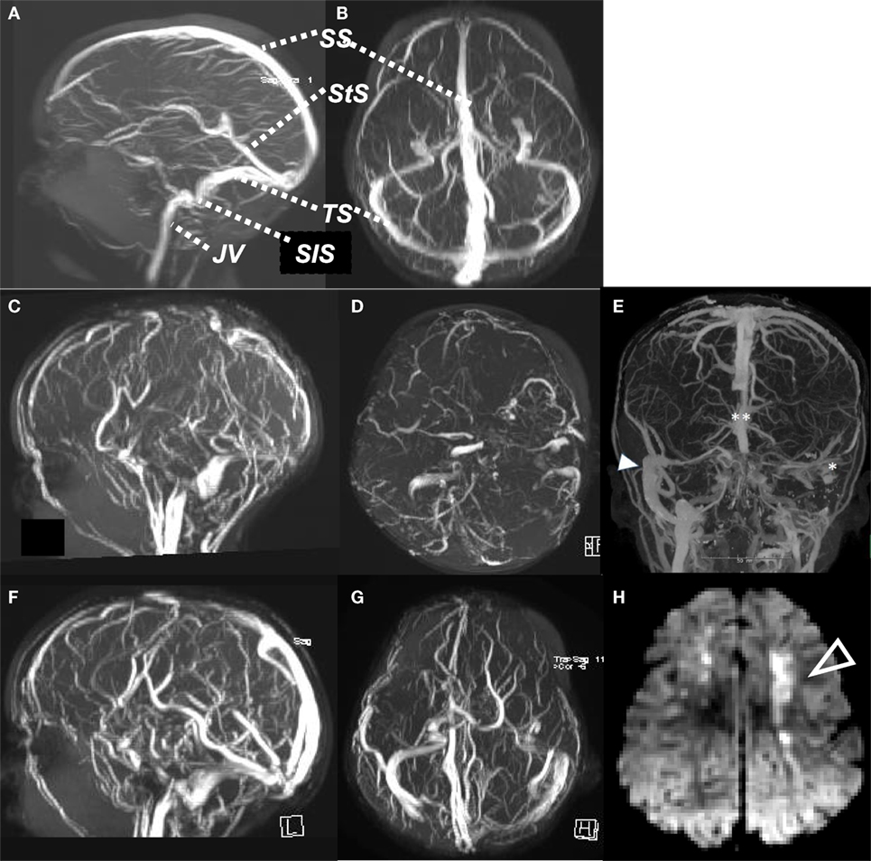


Frontiers Cerebral Sinovenous Thrombosis Pediatrics
Jan 29, 16 · Brain herniation with surrounding cerebrospinal fluid (CSF) into the dural venous sinuses is a uncommon entity and can simulate aforementioned pathologies and variations causing dural venous sinus filling It was recently described on magnetic resonance imaging (MRI) and it is also named as ''encephalocele" and "invagination"Dural venous sinus thrombosis (DVST) is a condition that ranges from being undiagnosed to leading to serious morbidity and mortality, including venous infarction and intracranial hemorrhage 1 DVST has a highly variable clinical presentation, from asymptomatic to acute or subacute headaches, signs or symptoms of increased intracranial pressure, focal neurologic deficits, orJun 17, 21 · Dural venous sinuses The human brain has the highest demand for oxygen of any tissue in the human body It receives a vast amount of blood from two systems the internal carotid system anteriorly, and the vertebrobasilar system posteriorly However, there is very little space in the cranial vault to accommodate large amounts of blood
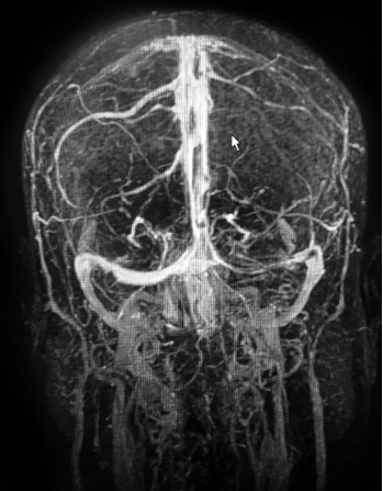


How To Spot And Treat Cerebral Venous Sinus Thrombosis Acep Now


Intracranial Venous System W Radiology
DVA or Developmental Venous Anomaly is pretty much what it sounds like It is a peculiar vein which goes back in time to when we were tiny little embryos, about a fifth of an inch (5mm) long Back then, when the brain was just beginning to form, its veins were very different from what they are now As we grew from a fraction of an inch to aDec 01, 14 · Cerebral venous thrombosis (CVT) is a potentially lifethreatening emergency The wide ranging of clinical symptoms makes the use of imaging in "slices" even more important for diagnosis Both CT and MRI are used to diagnose the occlusion of a venous sinus, but MRI is superior to CT for detecting a clot in the cortical or deep veinsCerebral venous thrombosis is often associated with nonspecific clinical complaints In addition, the imaging findings are often subtle Underdiagnosis or misdiagnosis of cerebral venous thrombosis can lead to severe consequences, including


Normal Structures In The Intracranial Dural Sinuses Delineation With 3d Contrast Enhanced Magnetization Prepared Rapid Acquisition Gradient Echo Imaging Sequence American Journal Of Neuroradiology
%20magnetic%20resonance%20venogram%20anatomy%20image%20sagiital.jpg)


Veins Of Brain Mrv Brain Veins Anatomy
Spiral brain CT demonstrated a hemorrhagic venous infarct in the right parietooccipital area associated with dense clot sign in the posterior part of the superior sagittal sinus (Fig 1 a) Brain MRI and MRV confirmed the diagnosis of CVST with displaying widespread thromboses in cortical veins, superior sagittal, transverse, and sigmoidOct 07, 19 · In venous occlusion, changes in the brain parenchyma can develop secondary to vasogenic edema, cytotoxic edema, or intracranial hemorrhage, giving rise to two possible scenarios CVT with local effects and CVT of the venous sinuses with increased intracranial pressure CVT leads to the formation of an area of focal cerebral edema because ofFeb 25, 18 · Cerebral venous thrombosis (venous sinus thrombosis) is caused by clots in the dural venous sinuses and accounts for 05% to 1% of all strokes CVT results in an increased venous pressure that can lower cerebral perfusion pressure and induce parenchymal change due to vasogenic edema, cytotoxic edema, or intracranial hemorrhage


Dural Venous Sinuses Radiology Reference Article Radiopaedia Org


Temporal Lobe Parenchyma Herniation Into The Transverse Sinus Mri Findings In A Case


Imaging Of Cerebral Venous Thrombosis Sciencedirect
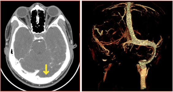


Annals Of B Pod Dural Venous Sinus Thrombosis Taming The Sru


Slow Flow V Thrombus Questions And Answers In Mri


Venous Sinuses Neuroangio Org


Cureus Cerebral Sinus Venous Thrombosis In The Setting Of Acute Mastoiditis
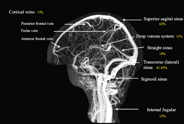


Cerebral Venous Thrombosis Core Em
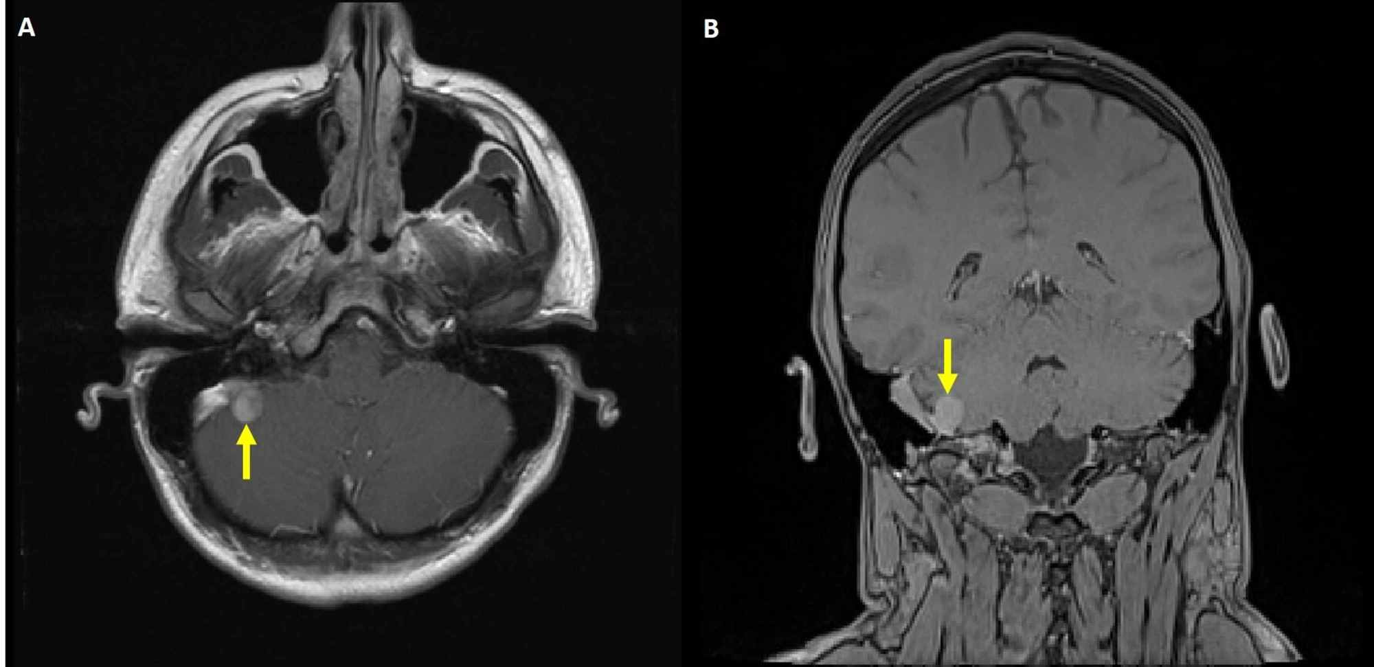


Cureus Venous Manometry As An Adjunct For Diagnosis And Multimodal Management Of Intracranial Hypertension Due To Meningioma Compressing Sigmoid Sinus


Cerebral Venous Sinus Thrombosis Wikipedia


A Mri Brain Showing Lack Of Flow Void Of Left Sigmoid Venous Sinus Download Scientific Diagram
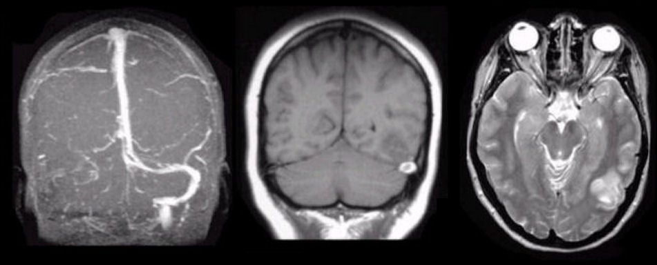


Sinus Thrombosis
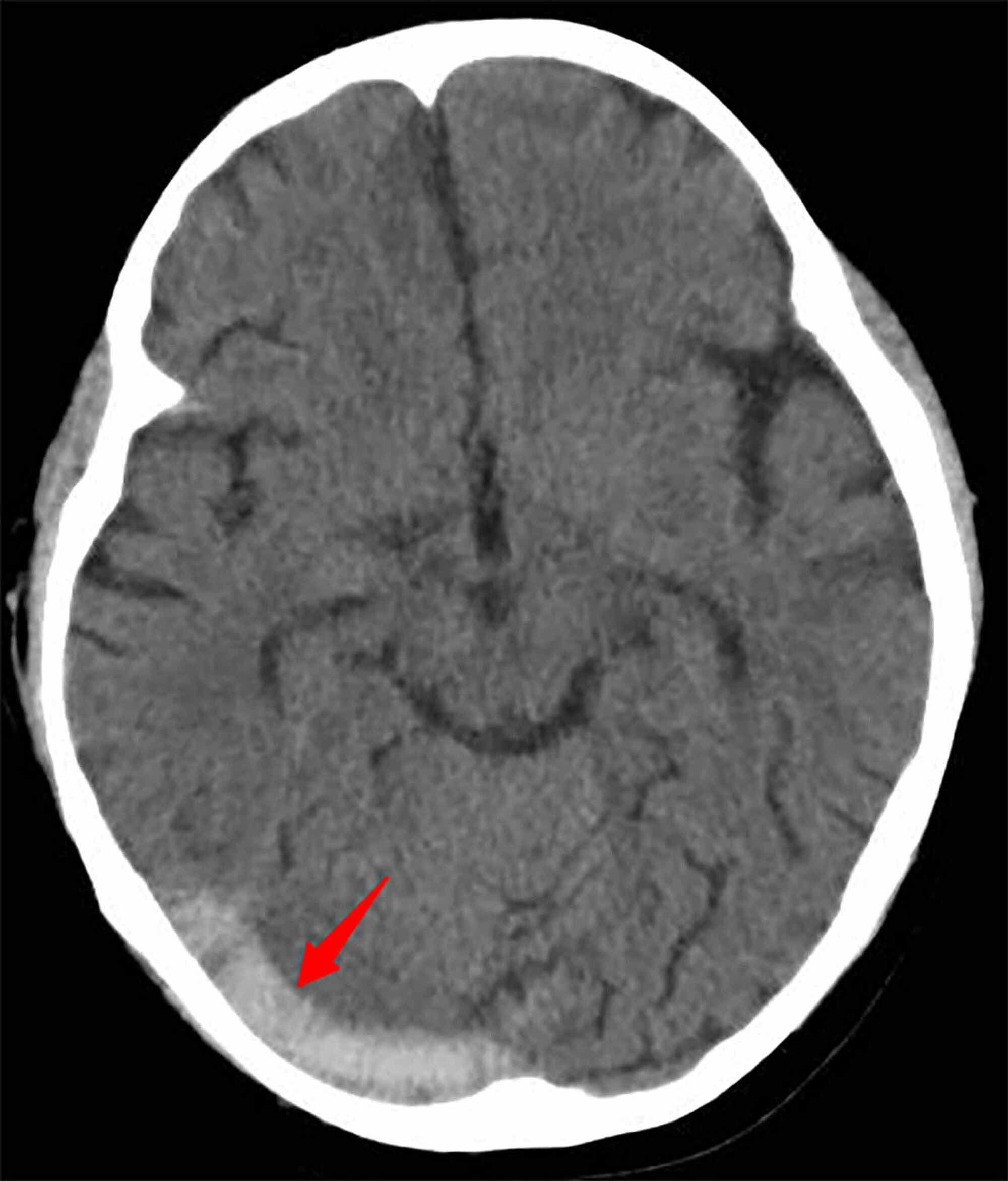


Cureus Cerebral Venous Sinus Thrombosis In A Child With Idiopathic Nephrotic Syndrome A Case Report And Review Of The Literature
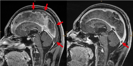


Cerebral Vein And Sinus Thrombosis


Venous Drainage Of The Cns Cerebrum Teachmeanatomy


Pin On Neurosurgery


Diagnostic Performance Of Routine Brain Mri Sequences For Dural Venous Sinus Thrombosis American Journal Of Neuroradiology


Dural Venous Sinus Thrombosis For Radiology Imaging


Focal Brain Herniation Into A Dural Venous Sinus An Incidental Rare Entity


Partially Recanalized Chronic Dural Sinus Thrombosis Findings On Mr Imaging Time Of Flight Mr Venography And Contrast Enhanced Mr Venography American Journal Of Neuroradiology


Imaging Of Cerebral Venous Thrombosis Sciencedirect


Brain Veins And Venous Sinuses Mri Angiogram Stock Image C048 9052 Science Photo Library


Radiological Imaging Of Cerebral Venous Thrombosis Ppt Video Online Download


Coronal T2 Brain Mri Revealing An Engorged Appearance Of The Bilateral Download Scientific Diagram


Imaging Of Cerebral Venous Thrombosis Clinical Radiology


Cerebral Sinus


Neuroanatomy Brain Veins Article


Dural Venous Sinuses An Overview Sciencedirect Topics


How To Treat Cerebral Venous And Sinus Thrombosis Coutinho 10 Journal Of Thrombosis And Haemostasis Wiley Online Library


A Review Of Extraaxial Developmental Venous Anomalies Of The Brain Involving Dural Venous Flow Or Sinuses Persistent Embryonic Sinuses Sinus Pericranii Venous Varices Or Aneurysmal Malformations And Enlarged Emissary Veins In Neurosurgical


Cerebral Venous Thrombosis Ischemia Cancer Therapy Advisor


1


Intracranial Venous System W Radiology


Mechanical And Structural Characterisation Of The Dural Venous Sinuses Scientific Reports


Acute Blindness Due To Cerebral Venous Sinus Thrombosis In A Child Postgraduate Medical Journal
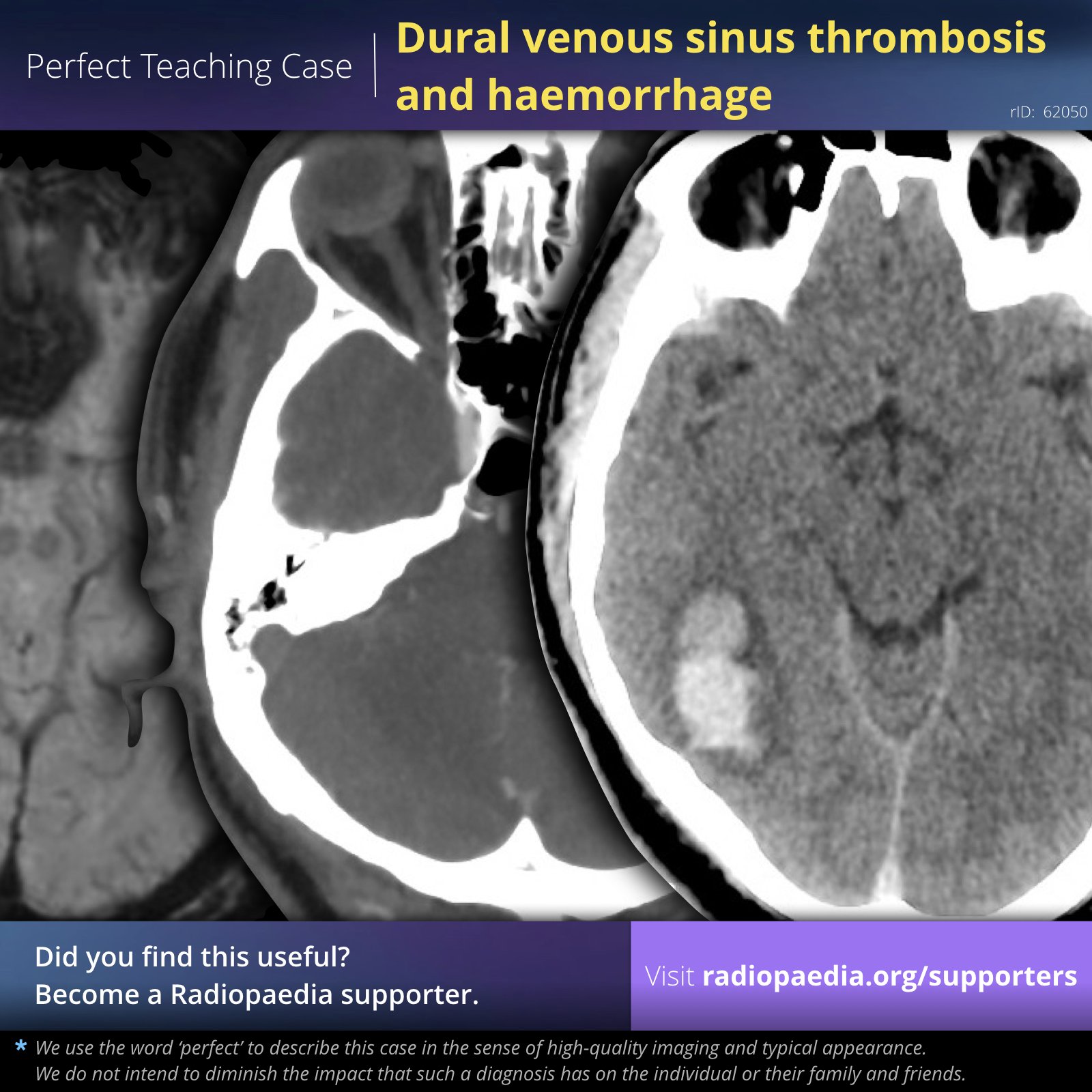


Frank Gaillard Perfect Teaching Case Dural Venous Sinus Thrombosis And Haemorrhage Mri And Ct View Case T Co Mczbyhi2mb Become A Radiopaedia Supporter T Co Uoz174knzo Supportradiopaedia Radiology Foamed Foamrad


Radiologic Clues To Cerebral Venous Thrombosis Radiographics


Influence Of Recanalization On Outcome In Dural Sinus Thrombosis Stroke


Emdocs Net Emergency Medicine Educationcerebral Venous Thrombosis Pearls And Pitfalls Emdocs Net Emergency Medicine Education


1


Venous Sinuses Neuroangio Org
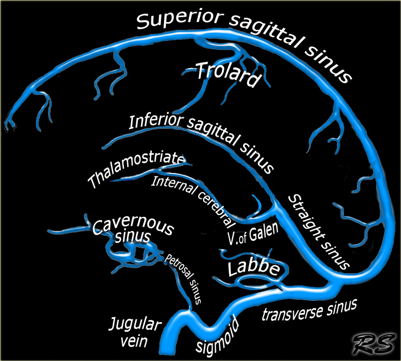


The Radiology Assistant Cerebral Venous Thrombosis


Cerebral Venous Thrombosis Epidemiology Diagnosis And Treatment Tidsskrift For Den Norske Legeforening


Cerebral Venous Thrombosis A Clinical Overview Intechopen


Mri Of Brain Venous Sinuses Study Youtube


Venous Sinus Thrombosis Radiology At St Vincent S University Hospital
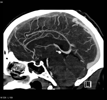


Dural Venous Sinus Thrombosis Radiology Reference Article Radiopaedia Org


Diagnostic Performance Of Routine Brain Mri Sequences For Dural Venous Sinus Thrombosis Ajnr News Digest


A Review Of Extraaxial Developmental Venous Anomalies Of The Brain Involving Dural Venous Flow Or Sinuses Persistent Embryonic Sinuses Sinus Pericranii Venous Varices Or Aneurysmal Malformations And Enlarged Emissary Veins In Neurosurgical


The Venous Distension Sign A Diagnostic Sign Of Intracranial Hypotension At Mr Imaging Of The Brain American Journal Of Neuroradiology
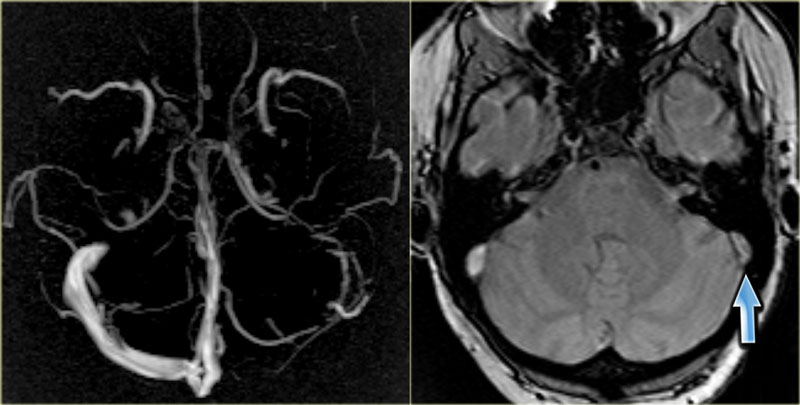


The Radiology Assistant Cerebral Venous Thrombosis
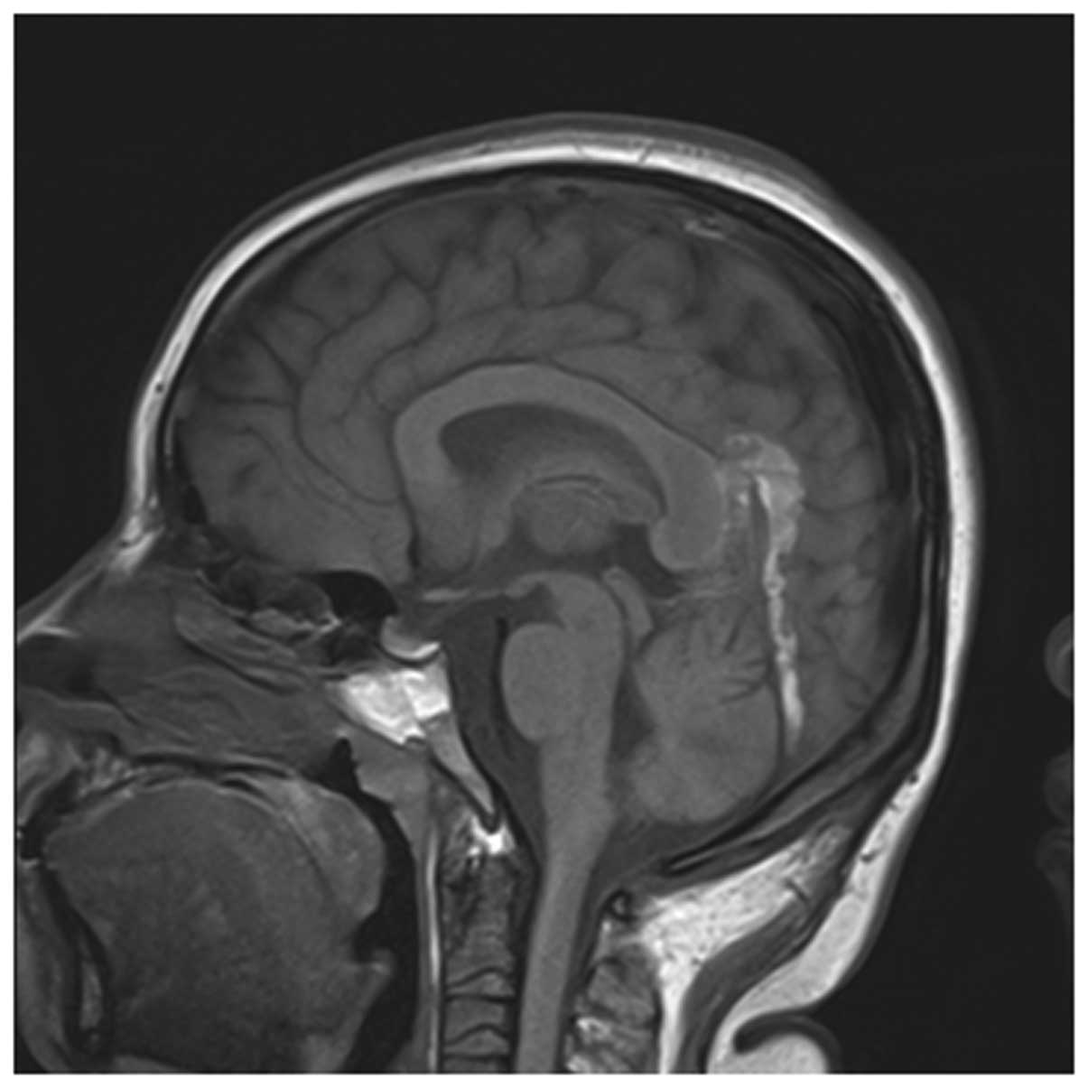


Early Imaging Characteristics Of 62 Cases Of Cerebral Venous Sinus Thrombosis


A T1 Weighted Mri The Superior Sagittal Sinus Shows A Hyper Intense Download Scientific Diagram


Venous Thrombosis Dominant Congenital Dysfibrinogenemia Presenting With Cerebral Venous Sinus Thrombosis Isth Congress Abstracts
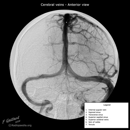


Transverse Sinus Radiology Reference Article Radiopaedia Org


Cerebral Venous Thrombosis Emcrit Project


Presentation1 Pptx Radiological Imaging Of Cerebral Venous Thrombosi


Intracranial Venous System W Radiology


Focal Brain Herniation Into A Dural Venous Sinus An Incidental Rare Entity
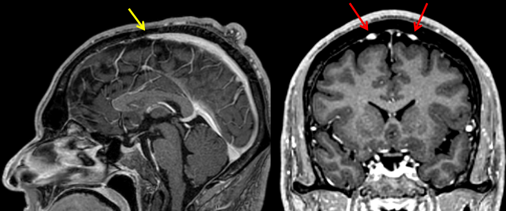


Epos C 0556
:background_color(FFFFFF):format(jpeg)/images/article/en/dural-sinuses/MBT9Z5mR2o8TnY2hb131w_Superficial_veins_of_the_brain_lateral_view.png)


Dural Venous Sinuses Anatomy Kenhub


Imaging Features Of Venous Sinus Thrombosis Part 2 Health4theworld Academy Youtube


Cerebral Venous Sinus Thrombosis Wikipedia


View Of Bilateral Transverse Sinus Hypoplasia A Rare Case Report Journal Of Experimental And Clinical Neurosciences


Venous Sinuses Neuroangio Org


Radiologic Clues To Cerebral Venous Thrombosis Radiographics
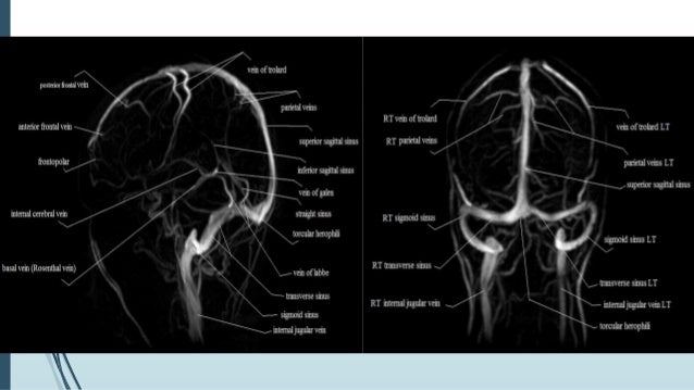


Dural Venous Sinus Thrombosis For Radiology Imaging
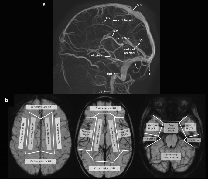


Venous Pathologies In Paediatric Neuroradiology From Foetal To Adolescent Life Springerlink


Dr Balaji Anvekar Frcr Hyperdense Dural Venous Sinuses On Ct


Dural Venous Sinuses 3d Anatomy Tutorial Youtube
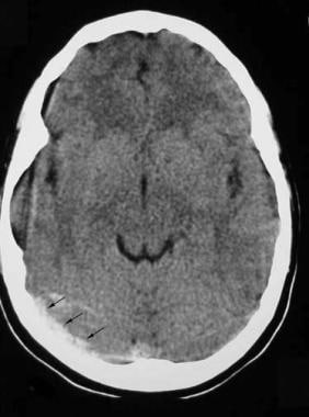


Brain Imaging In Venous Sinus Thrombosis Practice Essentials Computed Tomography Magnetic Resonance Imaging


Inferior Sagittal Sinus An Overview Sciencedirect Topics


Radiologic Clues To Cerebral Venous Thrombosis Radiographics


Cerebral Venous Sinus Thrombosis In Covid 19 Infection A Case Series And Review Of The Literature Journal Of Stroke And Cerebrovascular Diseases


Normal Structures In The Intracranial Dural Sinuses Delineation With 3d Contrast Enhanced Magnetization Prepared Rapid Acquisition Gradient Echo Imaging Sequence American Journal Of Neuroradiology


Teaching Neuroimages Congenital Variant Misdiagnosed As Cerebral Venous Sinus Thrombosis Neurology
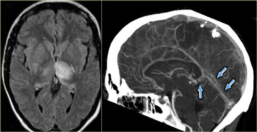


The Radiology Assistant Cerebral Venous Thrombosis


Figure 1 From Mri Diagnosis Of Dural Sinus Cortical Venous Thrombosis Immediate Post Contrast 3d Gre T1 Weighted Imaging Versus Unenhanced Mr Venography And Conventional Mr Sequences Semantic Scholar


Cerebral Venous Sinus Thrombosis Associated With Sars Cov 2 A Multinational Case Series Journal Of The Neurological Sciences
:background_color(FFFFFF):format(jpeg)/images/library/13677/8iVwiSwwNxvJhpSQELoQ_Sinus_sagittalis_superior_02.png)


Superior Sagittal Sinus Anatomy Tributaries Drainage Kenhub
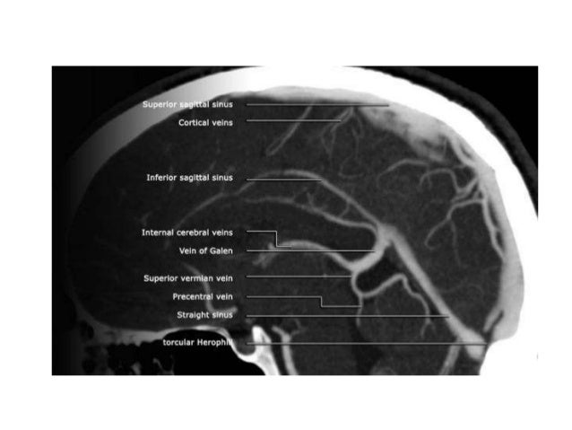


Venous Anatomy Of Brain Radiology


Cerebral Venous Sinus Thrombosis On Mri A Case Series Analysis


Brain Herniations Into The Dural Venous Sinuses Or Calvarium Mri Of A Recently Recognized Entity Semantic Scholar


Cerebral Venous Thrombosis A Diagnostic And Treatment Update Neurologia English Edition


コメント
コメントを投稿